






Organ transplants are arguably one of the most important human achievements in the field of medicine. The first ever successful heart transplant from a human donor to a human acceptor took place on December 3rd, 1967 (Brink & Hassoulas, 2009). The patient survived for about 18 days. A short but hopeful extension of life – and after decades of medical innovation, heart transplant patients today have a median survival of over 10 years (Khush et al, 2019).
According to the US Department of Health’s Division of Transplantation, over 3,000 adults and children are currently on the heart transplant wait list (Health Resources & Services Administration, 2024). Particularly for terminal patients, an organ transplant can be the last resort to helping extend a person’s life, particularly as a strong regimen of immunosuppressant medications have been developed to help prevent organ rejection.
But as scientists have monitored transplant patients, some have described a fascinating phenomenon: personality changes. Specifically, some transplant patients have reported experiencing the same emotions or feelings as their donors had (Al-Juhani et al, 2024). They may report suddenly having changes in personality traits, sexual preferences, or hobbies. In some cases, it’s simply a person expressing a change in their outlook on life, a reasonable and expected reaction after having almost come near death – but in other cases, actual changes in personality and preference appear that mirror donors.
Changes in Personality Following Heart Transplants
Personality changes can take shape in different ways such as changes in personal interests, foods, or even temperament. One Austrian study from 1992 looked at 47 heart transplant recipients, of which 3 reported having changes in their personality specifically due to their heart transplant (Bunzel et al, 1992). One patient reported a change in their temperament following their heart transplant, saying “the new heart has changed me … the person whose heart I got was a calm person, not hectic, and his feelings have been passed on to me now.”
In a compilation of case reports, Pearsall et al (2000) reported several examples; one woman, who had previously been highly athletic and healthy food-interested, started to crave Kentucky Fried Chicken, which her donor had loved. Another case described a middle-aged man, who received the heart of a young boy; despite previously disliking classical music, he began to love classical music, which the donor had played (Pearsall et al, 2000). One woman received the heart of a young girl who had been a vegetarian; despite previously being a meat-eater, the recipient could no longer stand eating meat and even experienced a change in her sexual preferences similar to the donor (Pearsall et al, 2000).
Even in a pediatric case, a 7-month-old infant received the heart of a 16-month-old infant and the recipient was reported to produce the same baby-talk words as the donor. The donor boy had also had a mild case of cerebral palsy that affected his left side; the recipient boy began to show some stiffness and shaking on his left side following the transplant (Pearsall et al, 2000). Overall, about 30% of heart transplant recipients exhibit some type of “ongoing or episodic emotional issues”
Cellular memory
As several studies have shown, heart transplant recipients experience various changes in temperament, preferences, and identity.
But how can a donor’s preferences and characteristics transfer over to the acceptor?
One 2020 paper hypothesized that within the transfer of a bodily organ, this phenomenon is caused by “cellular memory” (Liester, 2019). Memory is generally defined as a person’s ability to “acquire, store, and retrieve information”, with 3 general stages; acquisition, consolidation, and retrieval (Liester, 2019). Thus, cellular memory is based on the principle that various facets of a personality are biochemically stored within cells and are transferred to the recipient, whose own bodily mechanisms can retrieve such information “remember”. This can be done in various ways such as epigenetic memory, direct DNA memory, and RNA memory.
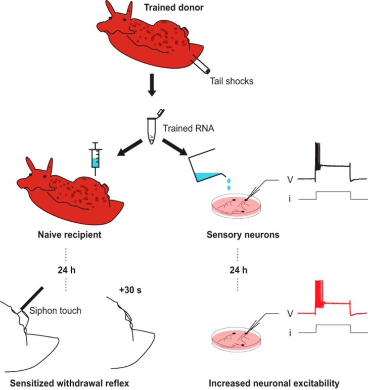
For example, several studies have found both epigenetic and RNA changes to be at play. In a 2018 study, scientists took memory-encoded cells from donor animals and injected into an acceptor animals led to some evidence of memory transmission. The research team used a mollusk species called Aplysia to study its effects. They trained “naïve” Aplysia by applying electric shocks to their tails until the molluscs learned to respond to the stimulus by withdrawing their tails, developing a long-term memory of the shock (Bédécarrats et al, 2018).
The molluscs had to encode the memory into their genetic code – but instead of directly changing their DNA code, they instead underwent a complex cellular process called DNA methylation, in which a chemical group made of a carbon atom bonded to three hydrogen atoms, was bonded to a portion of the DNA. DNA methylation has the effect of turning “off” specific regions of DNA and make them inaccessible. Whatever parts of the DNA remain “on” are then read by cell apparatus that transcribes the data into RNA. This leads to a significant change in how the DNA is read and transcribed, and this has subsequent effects on protein production and cell signalling. In other words, an experience caused the mollusc’s nerve cells to encode a memory and a subsequent response – withdrawal of the tail.
The scientists noted that the DNA methylation took place in these newly trained molluscs, signifying encoding, and they then isolated this RNA from their central nervous system. They injected it into other naïve untrained individuals, and they found that the acceptors started to experience significantly higher excitability signals in their sensory neurons and enhanced tail retraction behaviour compared to non-injected controls (Bédécarrats et al, 2018). They took on a feature of the donor, despite never having received the electric shock training themselves!
To further confirm these findings, the researchers then undid the effects of the DNA methylation by adding a DNA methylation inhibitor into half of the acceptor molluscs (Bédécarrats et al, 2018). The acceptors who had the inhibitor showed no significant excitability or tail retraction compared to the other acceptors. This further supported the fascinating way in which a simple stimulus could be encoded as a memory, stored, and then retrieved, even inside another organism!
While a human being is certainly not a mollusc, this pathway of cellular mechanisms is incredibly similar to our observations of new interests to certain sensory inputs in heart acceptors such as changes in their preferences for food, music, art, or sex.
Could such a similar phenomenon occur in humans? For many ethical reasons, this can be incredibly difficult to investigate in humans, and as such, no exact reason has yet been found, but the animal models have provided us with great insight.
How does memory transfer in humans?
So far, we’ve looked at theories on animal models such as molluscs, whose memories are stored within the simpler nervous system of the invertebrate animal.
But for humans, things are a lot more complicated. Our memories are usually stored in our brain, within our millions of synaptic connections.
So, how does cellular memory relate to the heart?
The heart is actually anchored within a complex network of nerves, including both nerves originating in the central nervous system, and nerve cells that are unique to the heart. The latter, also termed the “intracardiac nervous system” or ICNS , though functioning in the heart, is also capable of creating and storing memories (Figure 2).
During a heart transplant, the nerves of the ICNS are transplanted along with the heart. The ICNS is made up of approximately 200-1000 neurons, which are organized in combinations of clusters, called ganglionated plexi (GP) (Liester et al, 2019). These clusters of neurons receive signals and communicate signals, regarding various functions of the heart. The signals come in the form of neurotransmitters, such as norepinephrine, serotonin, histamine, dopamine, acetylcholine, and others, which are also all used in the brain. As a result, the heart is quite literally its own little mini brain, equipped with over 40,000 neurons which form a complex network (Al-Juhani et al, 2024)! This means that much of the fascinating phenomena in the brain, can also occur in the heart.
One interesting phenomenon is called “neuroplasticity.” Neuroplasticity is the ability of the nervous system to respond to changes in the environment by changing and adapting its structures and functions (Puderbaugh & Emmady, 2023). This is often seen following major changes to the brain, such as traumatic brain injury or stroke, but this can also be the case for the ICNS, following a major change such as the introduction of a foreign ICNS. Because the ICNS and the central nervous system are connected within the heart, it is theorized that the central nervous system of a heart recipient will connect with the ICNS of the donor and subsequently encode new information, especially when the heart is reinnervating within the acceptor’s body following the transplant.
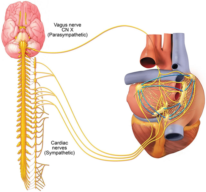
Since cardiac neurons may also encode memories as epigenetic modifications, these cells may most certainly will communicate with the brain. Researchers have found that about 80% of the signals between the brain and heart are ascending, meaning that most of the signals are transmitted from the heart up to the brain, which means that memories encoded from the donor could be communicated and transmitted into various parts of the brain, including the brain regions dealing with memories, fear, hormonal signals, movement, and even personality (Al-Juhani et al, 2024).
In one fascinating example, scientists determined that one major player in memory is prions. Prions are nonliving; they are not organisms so much as they are small proteins that fold in a certain way and then impose their own folding pattern on other proteins upon contact. This means that they can be infectious, and they do indeed play a major role in causing neurodegenerative diseases like mad cow disease, but they also have been implicated in memory storage. One example is a prion-like protein called cytoplasmic polyadenylation element-binding protein 3 or CPEB3. In certain physiological conditions, it folds in a certain way in 3D space, causing it to repress the activation of certain areas of RNA and reduce protein synthesis. However, scientists have found that during the creation of a new memory in the hippocampus, the CPEB3 prions fold into a different shape and aggregate into a unit that drives protein synthesis in neurons (Fioriti et al, 2015). When they inhibited the CPEB3, the long-term memory encoding was significantly reduced, further solidifying evidence that they play some major role in long term memory changes (Fioriti et al, 2015). Scientists have also noted that synapses can transport prions and proteins using small cellular packages called exosomes (Cheng et al, 2018), so this could be a method for the transfer of information between cells following a heart transplant.
It still remains a mystery to this day, but these theories may hold a great deal of potential answers. A heart transplant is a major life-saving operation, and it’s no wonder that it can often lead many to change their outlooks on their lives and ways of living – but there are instances of heart recipients who unquestionably acquire some of the same personality traits as their donors. The answers lie beyond the scope of our knowledge today, but we might just unlock the cellular basis of memory sooner than you think!
SOURCES
- Al-Juhani, A., Imran, M., Aljaili, Z. K., Alzhrani, M. M., Alsalman, R. A., Ahmed, M., Ali, D. K., Fallatah, M. I., Yousuf, H. M., & Dajani, L. M. (2024). Beyond the Pump: A Narrative Study Exploring Heart Memory. Cureus, 16(4), e59385. https://doi.org/10.7759/cureus.59385
- Bédécarrats, A., Chen, S., Pearce, K., Cai, D., & Glanzman, D. L. (2018). RNA from Trained Aplysia Can Induce an Epigenetic Engram for Long-Term Sensitization in Untrained Aplysia. eNeuro, 5(3), ENEURO.0038-18.2018. https://doi.org/10.1523/ENEURO.0038-18.2018
- Brink, J. G., & Hassoulas, J. (2009). The first human heart transplant and further advances in cardiac transplantation at Groote Schuur Hospital and the University of Cape Town - with reference to : the operation. A human cardiac transplant : an interim report of a successful operation performed at Groote Schuur Hospital, Cape Town. Cardiovascular journal of Africa, 20(1), 31–35.
- B. Bunzel, B. Schmidl-Mohl, A. Grundböck and G. Wollenek. (1992). Does Changing the Heart Mean Changing Personality? A Retrospective Inquiry on 47 Heart Transplant Patients. Quality of Life Research, 1(4), 251–256. doi:10.2307/4035054
- Cheng, L., Zhao, W., & Hill, A. F. (2018). Exosomes and their role in the intercellular trafficking of normal and disease associated prion proteins. Molecular aspects of medicine, 60, 62–68. https://doi.org/10.1016/j.mam.2017.11.011
- Fioriti, L., Myers, C., Huang, Y. Y., Li, X., Stephan, J. S., Trifilieff, P., Colnaghi, L., Kosmidis, S., Drisaldi, B., Pavlopoulos, E., & Kandel, E. R. (2015). The Persistence of Hippocampal-Based Memory Requires Protein Synthesis Mediated by the Prion-like Protein CPEB3. Neuron, 86(6), 1433–1448. https://doi.org/10.1016/j.neuron.2015.05.021
- Health Resources & Services Administration. 2024. Organ Donor Statistics, Detailed Description of Data. https://www.organdonor.gov/learn/organ-donation-statistics
- Khush, K. K., Cherikh, W. S., Chambers, D. C., Harhay, M. O., Hayes, D., Jr, Hsich, E., Meiser, B., Potena, L., Robinson, A., Rossano, J. W., Sadavarte, A., Singh, T. P., Zuckermann, A., Stehlik, J., & International Society for Heart and Lung Transplantation (2019). The International Thoracic Organ Transplant Registry of the International Society for Heart and Lung Transplantation: Thirty-sixth adult heart transplantation report - 2019; focus theme: Donor and recipient size match. The Journal of heart and lung transplantation : the official publication of the International Society for Heart Transplantation, 38(10), 1056–1066. https://doi.org/10.1016/j.healun.2019.08.004
- Liester, M. B. (2019). Personality Changes Following Heart Transplantation: The Role of Cellular Memory. Medical Hypotheses, 109468. doi:10.1016/j.mehy.2019.109468
- Mulpuru, S. K., & Shen, W. K. (2017). Selective modulation of the cardiac autonomic nervous system: a new strategy for treatment of cardioinhibitory syncope. Circulation: Arrhythmia and Electrophysiology, 10(2), e004994.
- Pearsall, P., Schwartz, G. E., & Russek, L. G. (2000). Changes in heart transplant recipients that parallel the personalities of their donors. Integrative medicine : integrating conventional and alternative medicine, 2(2), 65–72. https://doi.org/10.1016/s1096-2190(00)00013-5
- Puderbaugh M, Emmady PD. Neuroplasticity. [Updated 2023 May 1]. In: StatPearls [Internet]. Treasure Island (FL): StatPearls Publishing; 2025 Jan-. Available from: https://www.ncbi.nlm.nih.gov/books/NBK557811/